 |
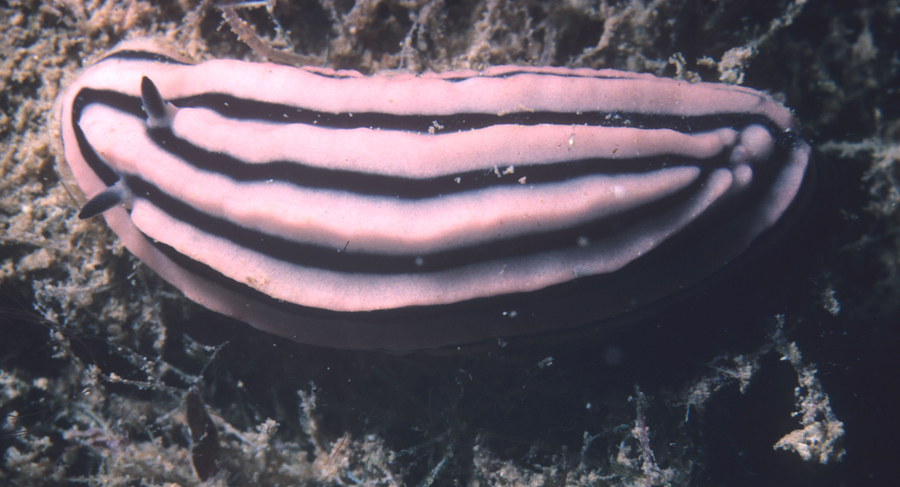
Phyllidiella rosans
In situ photograph of 28 mm long animal
14 October 1985, Papeete, Tahiti, 38 feet deep
Hans Bertsch photo
Phyllidiella rosans (Bergh, 1873)
This species was first named by Pease (1868), with the binomial Phyllidia nigrans. However, because of the prior use of this name, P. nigra van Hasselt, 1824, we must use the next available name for this species, Bergh's Phyllidia rosans. This shows the importance of maintaining a species' name with its author (P. nigra Pease is not P. nigra van Hasselt, but P. rosans Bergh!), and the use of parentheses around author and year to indicate placement of a species in a different genus than in which it was originally placed. Both species were named in Phyllidia Cuvier, 1797, but are now placed phylogenetically in Phyllidiella Bergh, 1869.
The body of Phyllidiella rosans is “oval with longitudinal pink rounded ridges. The ridges may be interrupted, especially in Pacific specimens. Anus opens at the apex of one of the posterior tubercles. Rhinophoral stalk pink and clavus black. Foot and gills grey” (Gosliner, Behrens & Valdés, 2008) . Each rhinophore on specimens > 30 mm has 12-15 lamellae. Live specimens range from 18-35 mm (Brunckhorst, 1993: 57). It occurs from Reunion and Oman to the Maldives in the western Indian Ocean, and from the Society, Marshall and Hawaiian Islands in the central Pacific.
There are at least 12 other pink and black phyllidiids in the Indo-Pacific (GBV, 2008: 292-295). They can be readily distinguished by color patterns, and presence or absence of ridges. Most characteristic is that the ridges of P. rosans are relatively smooth, lacking the obvious tubercles of the other species. The tubercles are most pronounced on the sides of the mantle .
Two distinctive morphological forms of P. rosans exist: “the 6–9 rounded, longitudinal ridges...may be interrupted in some individuals” (Brunckhorst, 1993: 57). They are both considered to be the same species (Brunckhorst, 1993, and GBV, 2008). Specimens collected by Andrew Garrett in Tahiti, and used by Pease (1868) and Bergh (1873) in their descriptions, were the uninterrupted ridge form. “Dorsum parte media varicositatibus tribus longitudinalibus continuis roseis pervagatum” (Bergh, 1873: 67). The synonyms Phyllidia soria Marcus & Marcus, 1970 (holotype: Mitirapa, Tahiti), and P. mediocris Yonow & Hayward, 1991 (I'Ile Maurice) match the uninterrupted-lined specimens illustrated by Pease and Bergh, whereas the longitudinal ridges of Plyllidia bourgini Risbec, 1928 (Citron Bay, New Caledonia), were interrupted. I have collected uninterrupted-line morphs in Tahiti (original locality of the Pease, Bergh and Marcus & Marcus descriptions), and the interrupted form on Oahu, Hawaii .
Numerous photographs of both morphological forms from across the Indo-Pacific are posted on the internet. Many can be accessed on Bill Rudman's Sea Slug Forum: www.seaslugforum.net and on links provided by this Slug Site. It would be most interesting to carefully analyze their geographic distribution, to determine whether allopatry exists between these morphs. This seems reminiscent of the patterns exhibited by the eastern Pacific Polycera alabe Collier & Farmer, 1964, and Limacia cockerelli (MacFarland, 1905).
The evolutionary significance of the foudroyant coloration of sea slugs has been a topic of concern for years. Two excellent recent summaries are Gosliner & Behrens (1990) and Terry Gosliner's “ color evolution in Nudibranchs .” “ Several passages in Dave Behrens' NUDIBRANCH BEHAVIOR (2005) summarize these reflections: “Their ability to evolve colors that blend with the background, a survival strategy known as cryptic or disruptive coloration, works extremely well for many species. Camouflage coats often come in an assortment of bland earth tones and greens that tend to match bottom environments....
“Not all cryptic species wear dull coloration. Several species of the dorid cryptobranchs camouflage by matching the vivid colors of their encrusting sponge prey....
“A number of cryptobranch dorids and a few sidegill slugs in family Pleurobranchidae derive protecton by duplicating not only their sponge preys' colors, but their textures as well....
“The aposematic coloration hypothesis has been successfully tested with nudibranchs in a number of studies. Some of these studies were simple field experiments where biologists tossed brightly colored nudibranchs into the water column in the presence of predatory fishes. In each case, fishes immediately approached the water-borne slug, and either eyed it closely and left it alone, or more commonly, engulf the slug and immediately spat it out. In the first case, the fishes had learned from previous encounters [sic] that these brightly colored critters were unpalatable. The second group of fishes were probably experiencing their first or second unpleasant encounter with the nudibranch.
“Controlled laboratory aquarium studies have demonstrated [the s]ame as in the field studies, some of the fishes, apparently reacting to the bright warning colors, made no attempt to eat the nudibranchs” (pp. 142, 146 and 150).
Having spent many hours UW in my búsqueda para nudibranquios (i.e., 'branching), I have come to realize that colors are not always what they seem. And we haven't even begun to explore the UV wavelengths! Some years ago (Bertsch, 1988), I wrote, “Observations in situ on the coloration of Phyllidia rosans Bergh, 1873, provide evidence on the adaptive significance of nudibranch colors. Most descriptions of nudibranchs are based on living animals in aquaria or in photographs made with a bright flash. Colors are usually vivid, leading to erroneous statements that these animals have warning coloration. The vast majority of nudibranchs under rocks or at subtidal depths blend in with their surroundings. Their bright colors are apparently energetically cheap methods by which the animal blends in with its variegated and muted surroundings.” Shallow-water tropical zones are an obvious exception. Phylogenetic studies significantly help us understand the evolutionary patterns of these biochemical features.
This animal shows that the differential absorption of light's wavelengths by water results in different colors visible (at least to us!). “In aquaria, Phyllidia rosans has alternating light pink and black longitudinal lines on its dorsum. However, on the back reef at Papeete, Tahiti, in situ on a cliff face at 13 m depth, P. rosans appeared bluish and black, blending in with its environment. Another specimen found in the same habitat on a bluish sponge at 21 m depth appeared to my vision with the natural lighting the same color as did the sponge. The pinkish color seen on the dorsum with a flash or in an aquarium was a muted bluish tone in the natural habitat” (Bertsch, 1988).
Many species of Phyllidiidae “have a characteristic odor derived from sesquiterpene isocyanides that the nudibranchs obtain from their prey and store in their mantle for defense. Phyllidiid chemicals are extremely toxic and can kill other organisms kept in aquaria together with the nudibranchs” (GBV, 2008: 284). “Stinkiness” may have been reported for the first time by Pease (1868: 80), when he wrote, “Emits a fetid odor.”
Literature Cited
Behrens, David W. 2005. Nudibranch Behavior. New World Publications, Inc., Jacksonville, Florida. 176 pp.
Bergh, L.S.R. 1873. Neue Nacktschnecken der Südsee, malacologische Untersuchungen. Journal des Museum Godeffroy 1(2): 65-96.
Bertsch, Hans. 1988. A review of Tahitian opisthobranchs: Faunal diversity and observations on color. Western Society of Malacologists, Annual Report 20: 25.
Brunckhorst, David J. 1993. The systematics and phylogeny of phyllidiid nudibranchs (Doridoidea). Records of the Australian Museum, Supplement 16: 1-107.
Gosliner, Terrence M. & David W. Behrens. 1990. Special resemblance, aposomatic coloration and mimicry in opisthobranch gastropods. In: Wicksten, M. (compiler), Adaptive Coloration in Invertebrates. Texas A&M University, College Station, Texas. pp. 127-138.
Gosliner, Terrence M., David W. Behrens & Ángel Valdés. 2008. Indo-Pacific Nudibranchs and Sea Slugs. A field guide to the World's most diverse fauna. Sea Challengers Natural History Books, Etc., Gig Harbor, WA, and California Academy of Sciences, San Francisco. 426 pp.
Pease, William Harper. 1868. Descriptions of marine Gasteropodae, inhabiting Polynesia. American Journal of Conchology 4 (2): 71-80.
Hans Bertsch
Universidad Autónoma de Baja California Sur, La Paz, BCS, México
and Imperial Beach, CA
]

|
