 |
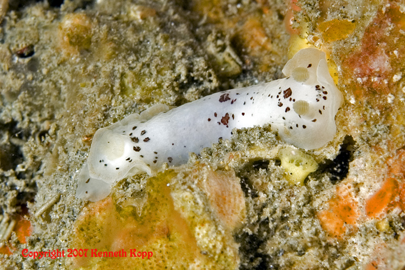
Dendrodoris behrensi
Photo courtesy of Kenneth KoppPhoto taken Cactus Cove, Catalina Island
Dendrodoris behrensi Millen & Bertsch 2005
There is nothing I enjoy more than writing about this great little critter. I wonder why. Since its description and presentation on this site , divers have been sending me sightings like mad. Ken’s photo here shows this species at a dead run, mantle extended and flapping. Gorgeous shot.
Immediately following Millen & Bertsch’s description is a paper by Jeff Goddard describing the very interesting larval development employed by this species. Specifically, Dendrodoris behrensi has ametamorphic direct development. Don’t ya just love some of these polynomial Anglo-Saxon derivatives.
While most species of nudibranchs lay eggs that hatch into planktonic “Veliger” larvae, this species does not. Instead, the embryos pass through the veliger stage lacking a shell and operculum. With the help of Jeff and the following microscopic photos he has generously contributed, let’s start at the beginning.
The egg mass of D. behrensi is a typical white coil just under 2 mm high, composed of a single spiral attached to the substrate by one edge. Each mass contains about 100 very large eggs . At about 24 days the embryos have well-developed eyespots, and can be observed rotating slowly within the egg capsule.
On day 38-ish, the embryos hatch as juvenile slugs, rather than a swimming veliger larvae. In Jeff’s side view you can see the eyespot (upper right) and other parts of its internal anatomy including kidney, hindgut, cerebral ganglion, and mouth parts. Looking down from above one can see the rhinophores (upper left in front of the eyespots), and the spicules which give the slugs body it structure and rigidity. When I was in college, we were told that the spicules found in the tissue of nudibranch bodies came from the sponges they fed upon. Not true. Of course my prof’s could not explain how species who fed on prey other than sponges had spicules also. Subsequently we have learned that these spicules develop beginning during the embryonic stage. These glass-like rodlets are secreted by a cell referred to as a scleroblast, and interestingly the spicule grows within this cell from its two ends toward the center.
Thanks guys!
REFERENCES:
Goddard, J.H.R. Ametamorphic direct development in Dendrodoris behrensi
(Nudibranchia: Dendrodorididae), with a review of developmental mode in the
family. Proceedings of the California Academy of Sciences 56: 201-211.
Millen, S.V. and Bertsch, H. (2005) Two new species of porostome nudibranchs
(family Dendrodorididae) from the coasts of California (USA) and Baja California (Mexico).
Proceedings of the California Academy of Sciences, 56(18):189-199.
Gig Harbor, Washington
Jan., 2007
Webmaster's Notes:
Once again Kenneth has demonstrated as with his photos of Hypselodoris californiensis , that sea slug photos are not a slam dunk as many underwater photographers might believe. Truly great slug shots are set aside by the forethought and intensity of the U/W photographer. We are all on a learning curve that seemingly has no end with the relentless advances in new technology. One common low tech denominator for all underwater photographers is the need to get wet to capture that picture of a life time!
Kenneth Kopp

The D. Behrensi was shot on the back side of Catalina at Cactus Cove (between Eagle Rock and the West End). Subject photograped with the Nikon rig described below, using a 105mm macro lens. This D. behrensi was walking solo at about 75 FSW in a cleft between two big rocks. It was tough to shoot as it was in there so deep. I have no idea how we saw it. I got really negative, and pressed my stomach against one rock while repositioning the strobes into the crack to illuminate it from both sides with enough light to shoot it at about 1/500 of a second. It was one of 6 different species ( Spanish Shawl, Limbaugh's, Yellow Edged Cadlina, MacFarland's, Mexichromis and this one) we (Claudette...my principal dive buddy and professional Nudi spotter and I) saw on that dive! We had no idea what it was until we got back to the boat and grabbed the new Nudi Card - on the dive I thought it was a flat worm because of that cow-catcher foot extension below its "head"... I didn't see the rhinophores or gill ring on the LCD underwater. I was so excited to find something new (to me, anyway...) Diving since 1999. Currently doing about 200 local dives a year. Favorite Nudibranch dive is Old Marine Land, Palos Verdes, CA. We routinely see 12 - 14 species on a single dive there.
|
I'm going to start shooting with a diopter soon - probably a 2X or 3X - so I can really get in tight. There is so much going on that is so tiny - Macro shots are the shots I love the best. So much life in a few square inches of our SoCal waters. I shoot hundreds of Nudi shots a month. Last year over my 254 dives, I probably shot over four thousand Nudi shots.
Writer and Marketing executive by profession. When I'm dry, I'm usually fly fishing or pounding and shaking things as a working drummer & percussionist.
Married, no kids. We have four house Rabbits. Yeah - its weird for me, too.
See more of Ken's work at bow_pix
Send Ken email at mo2vation@comcast.net
March 2005
Ali Hermosillo and Dave Behrens
Author:
Pacific Coast Nudibranchs and Nudibranch Behavior
Send Dave mail at dave@seachallengers.com
|
