 |
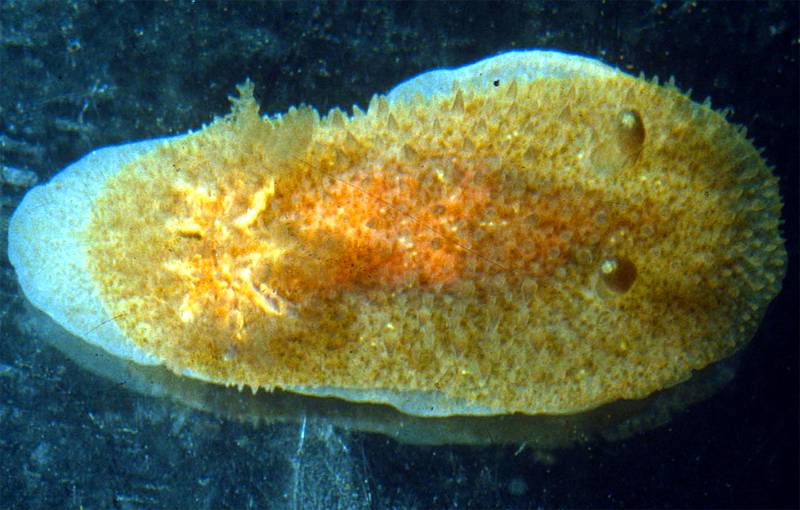
Acanthodoris planca
Photo courtesy of Shireen FaheyPhoto taken by Bill Liltveld
Cove Rock, False Bay, South Africa
Acanthodoris planca Fahey & Valdes, 2005
The body shape of Acanthodoris planca is oblong and the foot extends beyond the mantle margin. The dorsum is covered with elongate papillae that appear short and conical when preserved. The papillae in the preserved specimens are longer at the mantle margin. The oral tentacles are short and blunt. The rhinophores are stout clubs that angle towards the posterior and have 15–17 lamellae. The gill is broad and spreads to cover the posterior third of the animal. There are ten gill leaves that are bi- and tripinnate. The background color of the dorsum is pale brown or orange and there is a darker orange patch of coloration mid-dorsum. The foot is white with tiny brownish-orange dots. The rhinophores and gill leaves are the same color as the background color, but the rhinophore tips are white.Acanthodoris planca is externally most similar to the description of A. citrina Verrill, 1879, (a species that was later synonymized with A. pilosa by Thompson and Brown 1984) and to A. lutea MacFarland , 1925. All three species are orange yellow or orange and have a large gill that extends the width of the body. All three species have rhinophores that are similar in color as the body. The dorsum of each of these species is densely covered with conical tubercles. Acanthodoris planca has ten branchial leaves and both Verrill (1879) and MacFarland (1925) reported nine for both A. pilosa and A. lutea. The foot color differs between the species. Acanthodoris planca has a white foot with tiny brownish-orange spots, while that of A. lutea is orange yellow. Verrill did not report the foot color for A. citrina but the foot color of A. pilosa (Doris sparsa) was reported by Alder and Hancock (1846) as colorless and by Bergh (1879) as whitish or yellowish.
The radular morphology differs between these species. For example, Acanthodoris planca has densely arranged, multifid jaw rods, whereas A. lutea has triangular, hook-like structures. Verrill did not report the radular morphology of A. citrina but Thompson and Brown (1984) provided scanning electron micrographs of A. pilosa. Acanthodoris planca has 6–7 elongate, pointed outer lateral teeth, while A. lutea has 5–6 flattened, triangular plates “with a slight basal thickening” (MacFarland 1925). Acanthodoris pilosa has three outer lateral teeth that are flattened plates and a frail jaw cuticle with honeycombing (Thompson and Brown 1984). There are reproductive differences among all three species as well.
Citation :
Fahey, S. & Valdés, A. 2005. A review of Acanthodoris Gray, 1850 with a Phylogenetic Analysis of Onchiodorididae Alder and Hancock, 1845 (Mollusca, Nudibranchia), Proceedings of the California Academy of Sciences, 56:213-272.
San Francisco, Calif
Sep.. 2005
Photo courtesy of Dave Behrens
Taxonomic text courtesy of Shireen Fahey

Send Shireen email at sfahey@calacademy.org |
